

Bihar Biology HUMAN - Class 12 Bihar Board Zoology
Watch Video


Study smarter with Pi Lens
Get Notes, PYQs, Quiz and more for any Youtube video!
Bihar Biology HUMAN - Class 12 Bihar Board Zoology
62 views

PW Bihar Board English Medium
Notes
Video Summary
Human Reproduction
Definition and Core Concepts
Human reproduction is a biological process in which parents give birth to offspring through sexual reproduction, following viviparity. In humans, reproduction involves the formation and fusion of gametes, leading to the development of a new individual. Viviparity refers to direct birth of young ones, as opposed to oviparity, where offspring develop from eggs.
- Sexual reproduction: Involves the fusion of male and female gametes.
- Viviparity: Direct birth of offspring, characteristic of humans.
Male Reproductive System: Structure and Components
The male reproductive system consists of primary and secondary sex organs, each…
Human Reproduction
Definition and Core Concepts
Human reproduction is a biological process in which parents give birth to offspring through sexual reproduction, following viviparity. In humans, reproduction involves the formation and fusion of gametes, leading to the development of a new individual. Viviparity refers to direct birth of young ones, as opposed to oviparity, where offspring develop from eggs.
- Sexual reproduction: Involves the fusion of male and female gametes.
- Viviparity: Direct birth of offspring, characteristic of humans.
Male Reproductive System: Structure and Components
The male reproductive system consists of primary and secondary sex organs, each with specific roles in gamete production and hormone secretion. The primary sex organ is the testis, while secondary organs include accessory ducts, glands, and external genitalia.
- Primary sex organ: Testis (testes), responsible for sperm and hormone (androgen, mainly testosterone) production.
- Secondary sex organs:
- Accessory ducts: Rete testis, vasa efferentia, epididymis, vas deferens, ejaculatory duct.
- Accessory glands: Seminal vesicles (paired), prostate gland (single), bulbourethral glands (paired).
- External genitalia: Scrotum and penis.
Pathway of Sperm and Accessory Ducts
Sperm are produced in the seminiferous tubules and follow a specific pathway before ejaculation. The ducts facilitate transport, storage, and maturation of sperm.
- Pathway of sperm:
- Seminiferous tubules (site of sperm production)
- Rete testis
- Vasa efferentia
- Epididymis (site of sperm storage and maturation)
- Vas deferens
- Ejaculatory duct
- Urethra is not included in the sperm pathway as it serves as a common passage for both urine and semen.
Accessory Glands: Functions and Secretions
Accessory glands contribute fluids that support sperm viability, motility, and fertilization capability. Each gland has distinct secretions and roles.
- Seminal vesicles: Secrete fructose (energy source for sperm) and alkaline fluid; presence of fructose is unique to males and used in forensic tests.
- Prostate gland: Secretes milky fluid, provides citrate (energy), calcium (motility), and anti-coagulant factors (prevents semen coagulation).
- Bulbourethral (Cowper's) glands: Secrete lubricating fluid for the penis.
Testis: Structure and Cellular Organization
Testes are paired structures located in the scrotum, below the abdomen. Each testis contains compartments called testicular lobules, each with 1–3 seminiferous tubules.
- Dimensions: Length 4–5 cm, width 2–3 cm.
- Seminiferous tubules: Site of spermatogenesis; lined by two main cell types:
- Primary germ cells: Develop into sperm.
- Sertoli (nurse) cells: Provide nourishment and support to developing sperm.
- Interstitial (Leydig) cells: Located in interstitial spaces, produce androgens (mainly testosterone).
Spermatogenesis and Sperm Structure
Spermatogenesis is the process of sperm formation, starting from primary germ cells and involving mitosis and meiosis. Sperm structure is specialized for motility and fertilization.
- Stages of spermatogenesis:
- Primary germ cell → Spermatogonia (via mitosis)
- Spermatogonia → Primary spermatocyte (growth/differentiation)
- Primary spermatocyte (2n) → Secondary spermatocyte (n) (meiosis I)
- Secondary spermatocyte (n) → Spermatids (n) (meiosis II)
- Spermatids → Spermatozoa (spermiogenesis)
- Sperm structure:
- Head: Contains acrosome (modified Golgi body, contains hydrolytic enzymes), haploid nucleus.
- Neck: Contains centrioles (proximal and distal).
- Middle piece: Contains spirally arranged mitochondria (energy for motility).
- Tail: Flagellated, enables movement.
Semen and Seminal Plasma
Semen is the fluid ejaculated during copulation, consisting of sperm and seminal plasma (secretions from accessory glands). The composition and quality of semen are critical for fertility.
- Volume per ejaculation: 2–3 mL.
- Sperm count: Approximately 100 million per mL; 200–300 million per ejaculation.
- Fertility criteria:
- At least 60% of sperm must have normal shape and size.
- At least 40% must show vigorous motility.
Hormonal Regulation of Reproduction
Reproductive processes are regulated by hormones from the hypothalamus, pituitary, and gonads. The hypothalamus releases GnRH, stimulating the anterior pituitary to secrete FSH and LH.
- FSH (Follicle Stimulating Hormone): Acts on Sertoli cells, stimulates spermatogenesis.
- LH (Luteinizing Hormone/ICSH): Acts on Leydig cells, stimulates androgen (testosterone) production.
- In females:
- FSH stimulates follicle development and estrogen production.
- LH surge triggers ovulation and formation of corpus luteum (secretes progesterone).
Female Reproductive System: Structure and Oogenesis
The female reproductive system includes primary and secondary sex organs. Oogenesis is the process of ovum formation, characterized by emphasis on quality over quantity.
- Primary sex organ: Ovary (produces ova and ovarian hormones).
- Secondary sex organs:
- Accessory ducts: Vagina, cervix, uterus (all serve as passageways).
- Accessory gland: Mammary gland (milk production).
- External genitalia: Clitoris, labia majora, labia minora, mons pubis, hymen.
- Oogenesis:
- Begins during embryonic development; oogonia undergo mitosis to form primary oocytes.
- Primary oocytes arrest in prophase I (diplotene stage) until puberty.
- At puberty, meiosis I completes to form secondary oocyte and first polar body.
- Secondary oocyte arrests in metaphase II until fertilization.
- Only upon fertilization does meiosis II complete, forming the ovum and second polar body.
Ovarian Follicle Development and Ovulation
Ovarian follicles develop through several stages, each associated with specific cellular changes and hormonal influences.
- Stages:
- Primary follicle: Primary oocyte surrounded by a single layer of cells.
- Secondary follicle: Increased layers of follicular cells.
- Tertiary (antral) follicle: Formation of fluid-filled antrum.
- Graafian follicle: Mature follicle, contains secondary oocyte.
- Ovulation: Release of secondary oocyte from Graafian follicle into the fallopian tube, triggered by LH surge.
- Corpus luteum: Formed from the ruptured follicle, secretes progesterone.
Menstrual Cycle and Hormonal Events
The menstrual cycle is a 28-day cyclic event involving changes in the ovary and uterus, regulated by fluctuating hormone levels.
- Phases:
- Menstrual phase (days 1–5): Shedding of endometrial lining.
- Follicular/proliferative phase (days 6–14): FSH stimulates follicle growth; estrogen thickens endometrium.
- Ovulation (day 14): LH surge causes release of secondary oocyte.
- Luteal/secretory phase (days 15–28): Corpus luteum secretes progesterone, maintains endometrium.
- Key points:
- Estrogen: Responsible for secondary sexual characters and endometrial proliferation.
- Progesterone: Maintains secretory endometrium, essential for pregnancy.
- Ovulation day calculation: Cycle length minus 14 gives ovulation day.
- Expected delivery date: Last menstrual period + 7 days + 9 months.
Fertilization, Embryonic Development, and Placenta
Fertilization occurs in the ampulla region of the fallopian tube. The sperm penetrates the corona radiata and zona pellucida of the oocyte, triggering cortical reaction to prevent polyspermy.
- Fertilization steps:
- Sperm entry completes meiosis II in secondary oocyte.
- Formation of zygote, which undergoes cleavage to form morula (3–4 days), then blastula (6–7 days).
- Implantation of blastocyst in uterine wall (endometrium).
- Placenta:
- Functions: Nutrient and oxygen supply, waste removal, hormone production (hCG, hPL, estrogen, progesterone).
- Connects embryo to mother via umbilical cord.
- Acts as an endocrine tissue.
- Relaxin and inhibin are ovarian hormones, not placental.
Mammary Gland Structure and Milk Pathway
The mammary gland is responsible for milk production and secretion. The pathway of milk involves several structures, remembered by the mnemonic "Love Aaj Kal To Doosron Se Hota Hai, Apne To Drum Mein Daal Dete Hain, Neela Wala".
- Milk pathway:
- Mammary lobe → Alveoli → Mammary tubule → Mammary duct → Ampulla → Lactiferous duct → Nipple
- Colostrum: First yellowish milk, rich in IgA, provides immunity to the newborn.
- IgG: Only immunoglobulin that crosses the placenta.
Embryonic Layers and Organogenesis
After implantation, the inner cell mass of the blastocyst differentiates into three germ layers, each giving rise to specific tissues and organs.
- Ectoderm: Forms brain and nervous system.
- Endoderm: Forms digestive and respiratory systems.
- Mesoderm: Forms reproductive organs, connective tissues (bone, blood), muscles.
Gestation and Parturition
Gestation in humans lasts about 9 months, divided into three trimesters. Key developmental milestones occur in each trimester.
- First trimester (1–3 months): Heart forms first, followed by nervous system.
- Second trimester (4–6 months): Limbs, digits, eyelids, and hair develop.
- Third trimester (7–9 months): Complete organ maturation, preparation for birth.
- Parturition: Delivery of the baby, triggered by oxytocin and relaxin, involves uterine contractions.
- Lactation: Initiated post-delivery, colostrum is secreted for 6–7 days.
Key Exam Points and Common Questions
- Sertoli cells: Provide nourishment to developing sperm; controlled by FSH.
- Leydig cells: Produce testosterone; controlled by LH/ICSH.
- Sperm motility: Enabled by tail and mitochondria in the middle piece.
- Graafian follicle: Found in human ovary.
- Corona radiata: Group of cells surrounding the ovum.
- Zona pellucida: Protective membrane around the ovum.
- Polyspermy prevention: Achieved by cortical reaction at zona pellucida.
- Fertilization site: Ampulla region of fallopian tube.
- Implantation site: Endometrial lining of uterus.
- Birth canal: Formed by cervical canal and vaginal canal.
- Expected delivery date: Last menstrual period + 7 days + 9 months.
- Colostrum: Contains IgA, provides immunity to newborn.
- IgG: Only immunoglobulin crossing placenta to fetus.
This summary covers all major points, processes, and exam-relevant facts explicitly stated in the transcript.
Quiz
130 QuestionsClick on an option to find the answer
Question 1 of 130
In organisms with complex body designs, which of the following is TRUE about gametes?
Solution
The male gamete is motile, and the female gamete contains food stores
Observe the given image
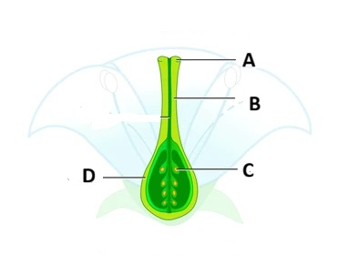
Match the correct label with its function.
(i) The part which accepts the pollen grain -
(ii) The part of the female reproductive system which carries egg cell in it -
(iii) The part which develops into fruit after fertilization-
Solution
(i)- (A) (ii)-(C) (iii) - (D)
Menstruation occurs if:
Solution
The egg is not fertilised
Flashcards
14 CardsClick to reveal answerTap the card to see answer

